A defining feature of eukaryotic cells is the presence of numerous membrane-bound organelles that subdivide the intracellular space into distinct compartments. Material exchange among most organelles occurs via vesicles that bud off from a source and specifically fuse with a target compartment. How the eukaryotic cell acquired its internal complexity is still poorly understood.
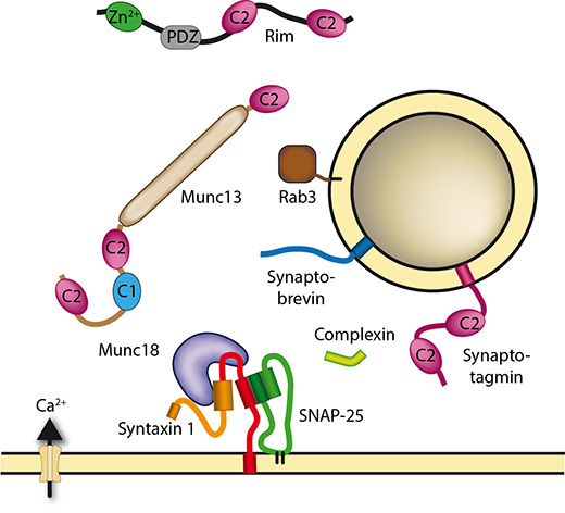
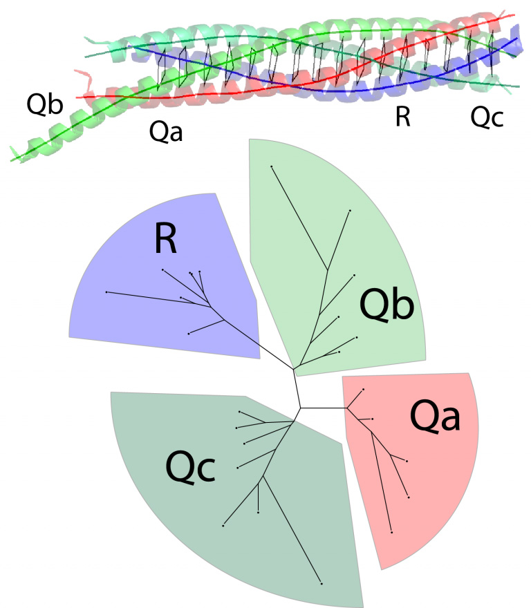
We strive to understand the mechanisms and the evolutionary history of the complex molecular machine that drives this important process. The key factors in vesicle fusion belong to conserved protein families. The core of the machine that drives the fusion of transport vesicles is composed of members of the SNARE protein family. They assemble into tight membrane-bridging complexes to pull two membranes together. Their activity is orchestrated by various other factors including Sec1/Munc18 (SM), Rab, and tethering proteins. Together, they form an ancient molecular machine that diversified during evolution to adapt to the needs of specialized compartments. Rapid exocytosis of neurotransmitters from synaptic vesicles constitutes one of such adaptations, and specific regulatory proteins such as synaptotagmins and complexins evolved in the animal kingdom.
Using biochemical, biophysical, morphological, and bioinformatics approaches we address the following questions:
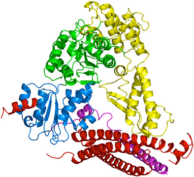
To better understand the molecular events underlying vesicular fusion we explore the physicochemical properties of the protein-protein interactions involved. We want to identify the domains involved, locate the binding surfaces and study the affinities, stoichiometries and kinetics of the interactions and their interplay with membranes to disentangle the entire network. For our biochemical studies, we almost exclusively use recombinant proteins. Next to standard biochemical techniques, we employ spectroscopic (circular dichroism and fluorescence spectroscopy) and calorimetric methods. Where feasible we use high-resolution structural techniques.
Concomitantly we want to shed light on the evolutionary history of the vesicle fusion machine, which arose from an ancient prototypic mechanism during the rise of the last eukaryotic common ancestor (LECA) from its prokaryotic-like ancestor. We would like to uncover how the mechanism adapted in different eukaryotic lineages and how it was most probably organized in the proto-eukaryotic cell. A particular focus lies on the evolutionary changes of the repertoire of the secretory machine during the rise of animals. Taking advantage of the huge number of available sequence data sets, our group developed a database to store and analyze sequences of the protein families involved in vesicle trafficking. Sequences are analyzed through iterative use of hidden Markov models and tree building.
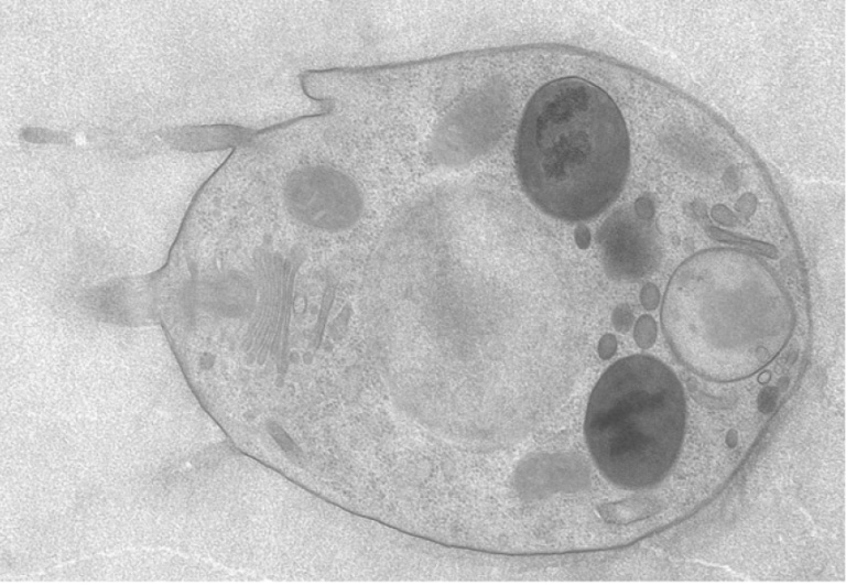
In conjunction, we plan to scrutinize in vivo, the interaction steps that we identified biochemically in vitro. Ultimately we want to correlate the configuration of the (neuro) secretory machinery in the cell with mutations/diseases-related to transport deficiencies. In addition, we are taking a closer look at the very early stages in the evolution of the secretory apparatus by studying the choanoflagellate Monosiga brevicollis and the placozoan Trichoplax adhaerens. Choanoflagellates are a group of mostly single-celled eukaryotes thought to be the closest known sister group to animals; Trichoplax is an animal positioned near the root of the animal tree. It is a simple, free-living marine animal without a nervous system and that glides using cilia to feed on algae. For this, we use, among others, state-of-the-art light and electron microscopy approaches.
Unveiling Conserved Allosteric Hot Spots in Protein Domains from Sequences.
bioRxiv 2024.05.13.593877
DOIExploring the conformational changes of the Munc18-1/syntaxin 1a complex.
Protein Sci. 2023;33(3):e4870
DOIArachidonic acid does not grease the exocytotic SNARE machinery.
bioRxiv 2023.02.06.527244;
DOI / bioRxivMegaviruses contain various genes encoding for eukaryotic vesicle trafficking factors.
Traffic. 2022 Aug; 23(8): 414–425
DOI / PMIDBET1 variants establish impaired vesicular transport as a cause for muscular dystrophy with epilepsy
EMBO Mol Med (2021)13:e13787
DOI / PMID / PDFHidden cell diversity in Placozoa: ultrastructural insights from Hoilungia hongkongensis.
Cell Tissue Res. 2021 Apr 19;10.1007/s00441-021-03459-y
DOI / PMID / PDFA conformational switch driven by phosphorylation regulates the activity of the evolutionarily conserved SNARE Ykt6.
Proc Natl Acad Sci U S A. 2021 Mar 23;118(12):e2016730118
DOI / PMIDChoanoflagellates and the ancestry of neurosecretory vesicles.
Philos Trans R Soc Lond B Biol Sci. 2021 Mar 29;376(1821):20190759
DOI / PMIDThe diversification and lineage-specific expansion of nitric oxide signaling in Placozoa: insights in the evolution of gaseous transmission.
Sci Rep. 2020 Aug 3;10(1):13020
DOI / PMIDPI(4,5)P2-dependent regulation of exocytosis by amisyn, the vertebrate-specific competitor of synaptobrevin 2
Proc Natl Acad Sci U S A. 2020 Jun 16;117(24):13468-13479
DOI / PMIDPrototypic SNARE Proteins Are Encoded in the Genomes of Heimdallarchaeota, Potentially Bridging the Gap between the Prokaryotes and Eukaryotes.
Curr Biol. 2020 Jul 6;30(13):2468-2480.e5
DOI / PMIDGlycine as a signaling molecule and chemoattractant in Trichoplax (Placozoa): insights into the early evolution of neurotransmitters.
Neuroreport. 2020 Apr 8;31(6):490-497
DOI / PMIDHigh Cell Diversity and Complex Peptidergic Signaling Underlie Placozoan Behavior.
Curr Biol. 2018 Nov 5;28(21):3495-3501.e2
DOI / PMIDEvidence for a conserved inhibitory binding mode between the membrane fusion assembly factors Munc18 and syntaxin in animals.
J Biol Chem. 292(50):20449-60
DOI / PMIDFunctional assays for the assessment of the pathogenicity of variants of GOSR2, an ER-to-Golgi SNARE involved in progressive myoclonus epilepsies.
Dis Model Mech. 10(12):1391-1398
DOI / PMIDGetting Nervous: An Evolutionary Overhaul for Communication.
Annu Rev Genet. 51:455-476
DOI / PMID / ServalShedding light on the expansion and diversification of the Cdc48 protein family during the rise of the eukaryotic cell.
BMC Evol Biol. Oct 18;16(1):215
DOI / PMID / AAA DatabaseThe SM protein Sly1 accelerates assembly of the ER–Golgi SNARE complex.
PNAS 111:13828-33
DOI / PMIDNovel cell types, neurosecretory cells, and body plan of the early-diverging metazoan Trichoplax adhaerens.
Curr Biol. 24:1565-72. (* co-last authors)
DOI / PMID / UNIL News / Comment in DOI / PMIDSyntaxin1a variants lacking an N-peptide or bearing the LE mutation bind to Munc18a in a closed conformation.
PNAS. 110:12637-42
DOI / PMIDPhosphatidylinositol 4,5-bisphosphate clusters act as molecular beacons for vesicle recruitment.
Nat Struct Mol Biol. 20:679-86
DOI / PMIDUntangling the evolution of Rab G proteins: implications of a comprehensive genomic analysis.
BMC Biol. 10:71
DOI / PMID / Rab DatabaseMunc18-1 mutations that strongly impair SNARE-complex binding support normal synaptic transmission.
EMBO J. 31(9):2156-68 (1: equally contributing authors; 2: corresponding authors)
DOI / PMIDPrimordial neurosecretory apparatus identified in the choanoflagellate Monosiga brevicollis.
PNAS. 108(37):15264-9
DOI / PMID / New ScientistA coiled-coil trigger site is essential for rapid binding of synaptobrevin to the SNARE acceptor complex.
J Biol Chem. 285:21549-59
DOI / PMIDSynaptobrevin N-terminally bound to syntaxin-SNAP-25 defines the primed vesicle state in regulated exocytosis.
Cell Biol. 188(3):401-13
DOI / PMIDA conserved membrane attachment site in α-SNAP facilitates NSF-driven SNARE complex disassembly.
J Biol Chem. 284(46):31817-26
DOI / PMIDSingle vesicle millisecond fusion kinetics reveals number of SNARE complexes optimal for fast SNARE-mediated membrane fusion.
J Biol Chem. 284(46):32158-66
DOI / PMIDThe Ca2+ affinity of synapto-tagmin 1 is markedly increased by a specific interaction of its C2B domain with phosphatidylinositol 4,5-bisphosphate.
J Biol Chem. 284:25749-60
DOI / PMIDIs assembly of the SNARE complex enough to fuel membrane fusion?
J Biol Chem. 284:13143-52
DOI / PMID / Comment in DOI / PMIDPhylogeny of the SNARE vesicle fusion machinery yields insights into the conservation of the secretory pathway in fungi.
BMC Evolutionary Biology 9:19
DOI / PMID / SNARE DatabaseImaging the assembly and disassembly kinetics of cis-SNARE complexes on native plasma membranes.
FEBS Letters, 582:3563-8
DOI / PMIDSNAREing the basis of multicellularity: Consequences of protein family expansion during evolution.
Mol.Biol.Evol. 25:2055-68
DOI / PMID / SNARE DatabaseMunc18a controls SNARE assembly through its interaction with the syntaxin N-peptide.
EMBO J. 27: 923-33
DOI / PMIDBinding of α-SNAP to syntaxin 1 blocks SNARE-dependent exocytosis.
Mol. Biol. Cell 19:776-84
DOI / PMIDSynaptotagmin activates membrane fusion through a Ca2+-dependent trans interaction with phospholipids.
Nat. Struct. Mol. Biol. 14: 904-11
DOI / PMIDBudding insights on cell polarity (News & Views).
Nature Structural & Molecular Biology 14: 360-2
DOI / PMIDAn elaborate classification of SNARE proteins sheds light on the conservation of the eukaryotic endomembrane system.
Mol. Biol. Cell 18: 3463-71
DOI / PMID / InCytes / SNARE DatabaseDeterminants of Synaptobrevin regulation in Membranes.
Mol. Biol. Cell 18: 2037-46
DOI / PMID / InCytesEarly endosomal SNAREs form a structurally conserved SNARE complex and fuse liposomes with multiple topologies.
EMBO J. 26: 9-18
DOI / PMIDN- to C-terminal SNARE complex assembly promotes rapid membrane fusion.
Science 313: 673-6
DOI / PMIDIdentification of SNAP-47, a novel Qbc-SNARE with ubiquitous expression.
J. Biol. Chem. 281: 17076-83
DOI / PMIDSequential N- to C-terminal ‘zipping-up’ of the SNARE complex drives priming and fusion of secretory vesicles.
EMBO J. 25:955-66
DOI / PMIDAlternative Splicing of SNAP-25 Regulates Secretion through Nonconservative Substitutions in the SNARE Domain.
Mol. Biol. Cell 12: 5675-85
DOI / PMIDA structural basis for the inhibitory role of tomosyn in exocytosis.
J. Biol. Chem. 279: 47192-200 “JBC Paper of the week”
DOI / PMIDA transient N-terminal interaction of SNAP-25 and syntaxin nucleates SNARE assembly.
J. Biol. Chem. 279: 7613-21
DOI / PMIDSingle-molecule fluorescence resonance energy transfer reveals a dynamic equilibrium between closed and open conformations of syntaxin-1.
PNAS 100: 15516-21
DOI / PMIDStructural insights into the SNARE mechanism.
BBA - Molecular Cell Research. Special Issue: Membrane Fusion, 1641: 87-97
DOI / PMIDThe R-SNARE motif of tomosyn forms SNARE core complexes with syntaxin 1 and SNAP-25 and down-regulates exocytosis.
J. Biol. Chem. 278: 31159-66
DOI / PMIDCrystal structure of a complex between human spliceosomal cyclophilin H and a U4/U6 snRNP-60K peptide.
J. Mol. Biol., 331: 45-56
DOI / PMIDHabc Domain and SNARE Core Complex Are Connected by a Flexible Linker.
Biochemistry, 42: 4009-14
DOI / PMIDSNARE assembly and disassembly exhibit a pronounced hysteresis.
Nat. Struct. Biol. 9: 144-151
DOI / PMID / Commented in DOI / PMIDCrystal structure of the endosomal SNARE complex reveals common structural principles of all SNAREs.
Nat. Struct. Biol. 9: 107-111
DOI / PMIDRapid and selective binding to the synaptic SNARE complex suggests a modulatory role of complexins in neuroexocytosis.
J. Biol. Chem. 277: 7838-48
DOI / PMIDHomo- and heterooligomeric SNARE complexes studied by site-directed spin labeling.
J. Biol. Chem. 276: 13169-77
DOI / PMIDA SNARE complex mediating fusion of late endosomes defines conserved properties of SNARE structure and function.
EMBO J. 19:6453-64
DOI / PMIDSelective interaction of complexin with the neuronal SNARE complex: determination of the binding regions.
J. Biol. Chem. 275:19808-18
DOI / PMIDKinetics of Synaptotagmin Respones to Ca2+ and Assembly with the Core SNARE Complex onto Membranes.
Neuron 24: 363-76
DOI / PMIDConserved structural features of the synaptic fusion complex: SNARE proteins reclassified as Q- and R-SNAREs.
Proc. Natl. Acad. Sci. USA 95: 15781-86
DOI / PMIDCrystal structure of a SNARE complex involved in synaptic exocytosis at 2.4 Å resolution.
Nature 395: 347-353
DOI / PMID / Comment in DOI / PMIDIdentification of a minimal core of the synaptic SNARE-complex sufficient for reversible assembly and disassembly.
Biochemistry 37: 10345-55
DOI / PMIDStructural changes are associated with SNARE complex formation.
J. Biol. Chem. 272, 28036-41
DOI / PMIDA structural change occurs upon binding of syntaxin to SNAP-25.
J. Biol. Chem. 272: 4582-90
DOI / PMIDComparative genomics and the nature of placozoan species.
PLoS Biol. 2018 Jul 31;16(7):e2005359
DOI / PMIDLoss of Neuroligin3 specifically downregulates retinal GABAAα2 receptors without abolishing direction selectivity.
PLoS One. 2017 Jul 14;12(7):e0181011
DOI / PMID82nd International Titisee Conference held from October 25 to 29, 2000. Mechanisms of membrane fusion.
B.I.F. FUTURA Vol. 16, No. 1, 13-23 (Conference report)
Download PDFMechanismen intrazellulärer Membranfusion.
Biospektrum No. 1, 2731 (review article; german)
Download PDFDirk Fasshauer studied biology and received his doctorate degree from the University of Göttingen in 1994. He worked as a post-doctoral fellow at Yale University from 1995 to 1997 and then moved to the Max Planck Institute for Biophysical Chemistry in Göttingen and from 2002, headed the “Structural Biochemistry” Research Group in the Department of Neurobiology. In 2009 he joined the DNF as associate professor.
Aisima studied Chemical and Biological Engineering at Koc University, Turkey. In her doctoral thesis, she dealt with finding allosteric networks in proteins and nanobody modeling. In 2020, she started as a postdoc at the Barth Lab, EPFL, where she studied reprogramming and modeling signaling properties in de-novo proteins. She joined the group as a postdoc in 2022.
Carlos received a BSc in Biology from the University of the Balearic Islands in 2014. Then he completed his master in Bioinformatics in 2017 at University Pompeu Fabra in Barcelona. In 2021, he finished his PhD in Biomedical Sciences at the University of Bern, where he worked on NGS analysis of cancer cell lines and designs of CRISPR-Cas9 libraries. In October 2021, he joined the group as a Postdoc.
Christian studied Biochemistry at the University of Zurich. His PhD research focused on using NMR spectroscopy to investigate picosecond-to-nanosecond dynamics in a GPCR and to trace the evolution of a metalloprotein in snails. Following his PhD, he worked as a scientific staff member at the Zurich Water Works. He joined the group as a Postdoc in October 2024.
Iman obtained her B.Sc. in Biochemistry in June 2016 at the Mohamed Khidher University in Algeria.Then, she obtained her Master in Molecular biology, Immunology, and Microbiology Specialities at the Eotvos Lorand University, Hungary. She started her PhD thesis in November 2020.
Michela studied Molecular and Medical biotechnology and received her Master's degree in March 2020 at the University of Verona, Italy, with a bioinformatic thesis about RNA sequencing analysis on lymphoma cancer. She started her PhD in August 2020.
Sévan received a B.Sc. in Biology in 2021 and a M.Sc. in Medical Biology in 2023 from the University of Lausanne. After a short stay in the Knobloch’s lab where he learned the basics of cell culture on stem cells and a master thesis in the Hummler’s lab where he studied the role of a membrane-bound serine protease in mice kidney, Sévan started a Ph.D. in Life Sciences and joined Dirk Fasshauer’s lab in January 2024.
Arianna received her Master’s degree in Quantitative and Computational Biology from University of Trento (Italy) in March 2020. She worked as a Bioinformatician at Prinses Maxima Centrum (Utrecht, the Netherlands) and Institut Français de Bioinformatique (Marseille, France) with the research focus on NGS analysis and data quality control. She started her PhD in May 2024.
Deepak received his Master's degree in Bioinformatics from IIIT Hyderabad in 2013. He worked as a researcher at TRDDC (TCS Research) Pune from 2013 to 2020, with the research focus on NGS analysis, metagenomics and network biology. He started his PhD thesis in November 2020.
In the SNAREs section of the database you have the possibility to view our classified sequences (SNAREs -> View DB) or to de novo classify protein sequences (SNAREs -> Find Motif). If you choose to view our classified sequences, you find additional information for each of the fields on the ViewDB site by clicking the question mark behind the field. If you choose to de novo classify protein sequences you may paste any protein sequence into the text field of the Find Motif site and hit the Submit-button. The results will be displayed shortly after.
Snare DBIn the AAAs section of the database you have the possibility to view our classified sequences (AAAs -> View Database) or to de novo classify protein sequences (AAAs -> Scan Sequence against HMMs). If you choose to view our classified sequences, you find additional information for each of the fields on the ViewDB site by clicking the question mark behind the field. If you choose to de novo classify protein sequences you may paste any protein sequence into the text field of the Find Motif site and hit the Submit-button. The results will be displayed shortly after.
AAA DBIn the Rabs section of the database you have the possibility to view our classified sequences (Rabs -> View Database) or to de novo classify protein sequences (Rabs -> Scan Sequence against HMMs). If you choose to view our classified sequences, you find additional information for each of the fields on the ViewDB site by clicking the question mark behind the field. If you choose to de novo classify protein sequences you may paste any protein sequence into the text field of the Find Motif site and hit the Submit-button. The results will be displayed shortly after.
RAB DB
UNIL Génopode
CH-1015 Lausanne
Switzerland